Our research: case studies

New study to test personalised breast cancer screening
A new international trial will investigate whether a personalised breast cancer screening can help detect the disease sooner.
MyPeBS trial is taking place in 6 European countries and plans to involve 85,000 volunteers aged between 50 and 70 who have never had breast cancer before.
Currently those between the ages of 50-70 are invited for a routine NHS breast cancer screening by having a mammogram every three years. However, not everyone has the same breast cancer risk, other factors such as genetics, hormones, family history and breast density can put some in a higher risk category.
The MyPeBS study randomly assigns trial volunteers to follow either the standard NHS screening schedule or a personalised screening schedule according to their risk of breast cancer.
Professor Fiona Gilbert, professor of radiology at University of Cambridge and imaging theme lead for the NIHR Cambridge Biomedical Research Centre is leading the UK study. “This is an opportunity to take part in one of the largest studies so far into how we find early stage breast cancer. By taking a saliva sample and history from those selected on the trial, we can identify whether they are at higher or lower risk of developing breast cancer. Once we know this, we can tailor screening to their own personal needs.”
Cambridge University Hospitals NHS Foundation Trust (CUH), the Leeds Teaching Hospitals NHS Trust and Manchester University NHS Foundation Trust (MFT) will be supporting the UK arm of the study and plans to recruit 10,000 volunteers which will last for 4 years. The Cambridge site will be supported by the NIHR Cambridge BRC.
For more information about the trail, go to: www.mypebs.eu
ITV News reported on the trial in October 2021 or read the full news article.

World first for AI and machine learning to treat Covid patients worldwide
In a ground-breaking study supported by the NIHR Cambridge BRC, Addenbrooke’s Hospital, healthcare technology firm NVIDIA and 20 other hospitals worldwide have used artificial intelligence (AI) to predict Covid patients’ oxygen needs.
In what’s known as federated learning, the research applied an algorithm to analyse anonymised electronic patient health data and chest x-rays from 10,000 Covid patients worldwide, including 250 at Addenbrooke’s Hospital.
The study – dubbed EXAM – took just two weeks of AI ‘learning’ to achieve high-quality predictions on how much extra oxygen a patient would need in the first days of hospital care.
To maintain strict patient confidentiality, the patient data was fully anonymised and an algorithm was sent to each hospital so no data was shared or left its location.
Once the algorithm had ‘learned’ from the data, the analysis was brought together to build an AI tool which could predict the oxygen needs of hospital Covid patients anywhere in the world.
This model can be used to help frontline physicians worldwide.
This is an abridged version of the article first published on our website on 15 September 2021.

Well-planned patient preparation before their scans improves imaging quality and cancer detection rates
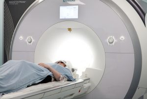
An NIHR Cambridge BRC-supported review of existing studies has found that adequate preparation of patients with suspected prostate cancer before their MRI scans could significantly improve imaging quality, cancer detection rates and subsequent treatment planning.
Multiparametric magnetic resonance imaging (mpMRI) is a special type of scan that creates more detailed pictures of the prostate. It plays an essential role in the diagnosis of prostate cancer (PCa), and can help doctors decide if tumours are clinically significant (that is, likely to impact on a patient’s life and therefore needing treatment); it is now performed in up to 75% of men with suspicion of PCa in the UK.
Existing guidelines on its use, while they address technical hardware and software and diagnostic considerations, have not to date adequately covered patient-related factors which also can affect the quality of the imaging.
This review from Cambridge radiologists Dr Tristan Barrett and Dr Iztok Caglič looked at how some simple steps can help to further optimise the image quality of mpMRI.
Before mpMRI the authors therefore recommend:
- Using antiperistaltic agents to minimise spasm in the intestines and associated movement (which can result in blurring of image)
- Asking patients to empty their bowels prior to examination to minimise image distortion
- Asking patients to refrain from ejaculation for three days before their scans
- Using an additional scan sequence called PROPELLER to improve image quality in patients with hip metalwork.
Read the full article: https://doi.org/10.1016/j.crad.2018.12.003

Prostate Cancer Diagnostic Pathways
After skin cancer, prostate cancer is the most common cancer in men, usually affecting those over 50. Researchers in Cambridge have led on MR imaging to better identify tumours within the prostate, coupled with precision biopsy techniques using fusion software.
Clinicians at Cambridge University Hospitals have been working with imaging researchers to carry out Magnetic Resonance Imaging (MRI) targeted prostate biopsies (needles into the prostate to retrieve samples), using MRI-Ultrasound image fusion software.
In 2011, Cambridge was the first centre in the UK to use this technique routinely in the clinical setting, for repeat biopsy in high-risk patients. This practice was subsequently adopted in the 2014 update of the NICE guidelines for prostate cancer. Cambridge researchers developed the Ginsburg group guidelines on how to perform targeted ‘transperineal biopsy’ to allow standardisation of the technique.
The traditional diagnostic pathway of prostate cancer has been changed since 2015; prior to intervention, men now have the MR imaging before undergoing a biopsy.
This pathway has provided clinicians an improved way to identify cancerous tumours in the prostate and has reduced the number of invasive samples being taken and in some cases avoiding a biopsy altogether.
This has allowed clinicians to “get it right first time” and is helping men to be diagnosed faster and start treatment earlier. The Anglian Network Cancer Group has adopted this practice as its Prostate Best Practice Pathway, and NICE have suggested that this practice will form part of their updated 2018 guidelines for prostate cancer diagnosis and management.

