Imaging


This theme’s focus:
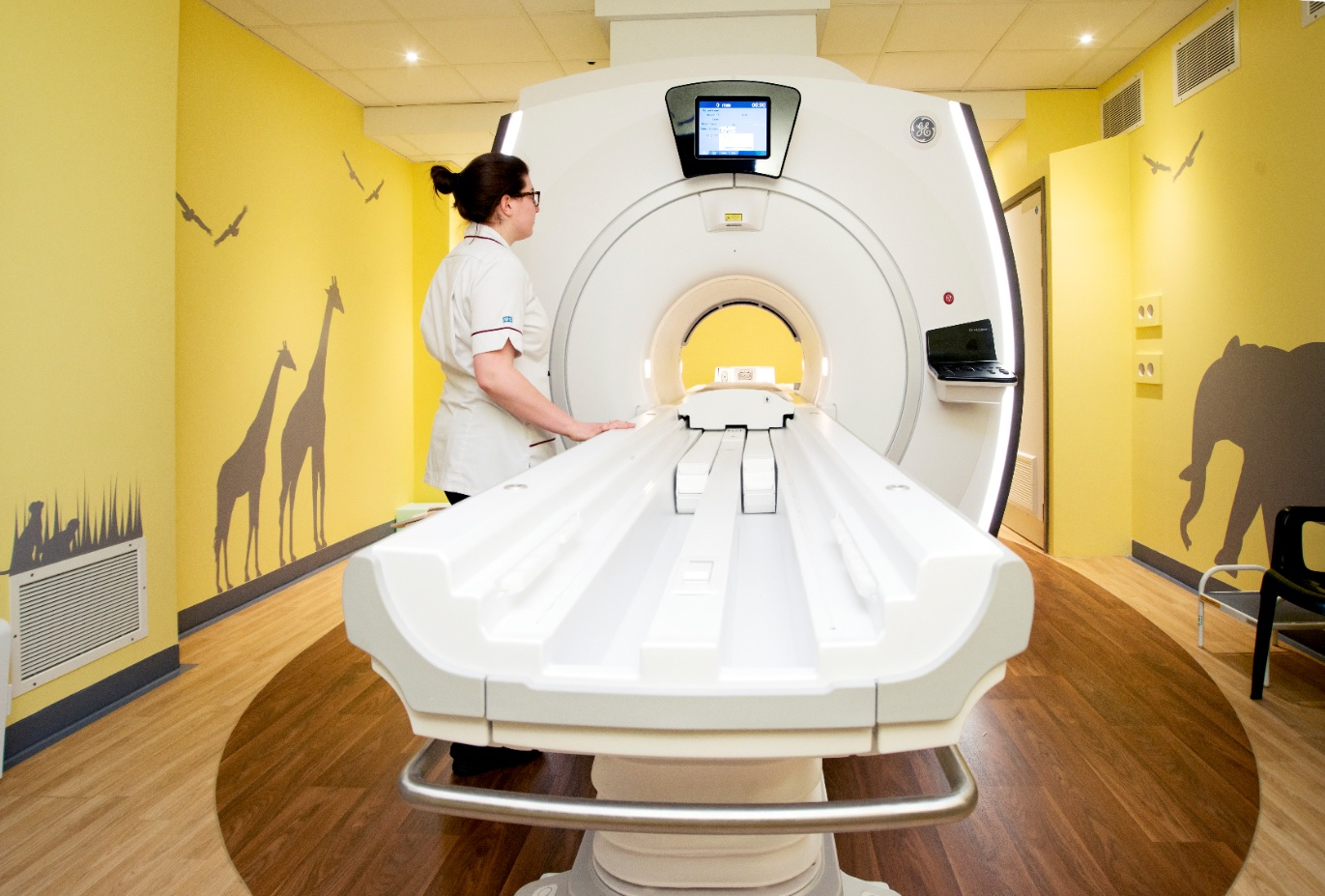
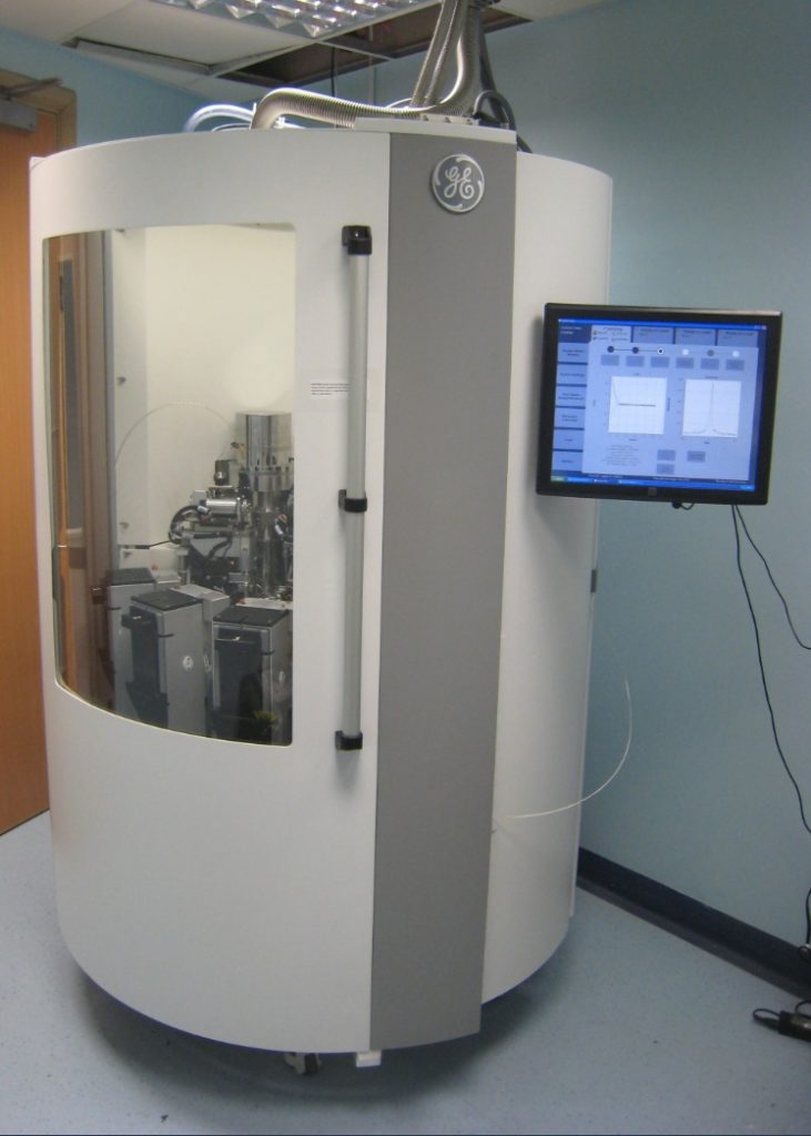
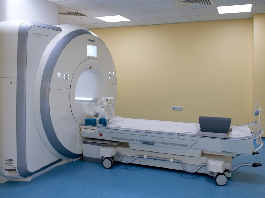
- Developing new, more sensitive imaging methods, especially for MRI (magnetic resonance imaging) and PET (positron emission tomography)
- Creating and testing new AI (artificial intelligence) techniques for better imaging and disease detection
- Translating these new methods into patient care through collaborations across the UK and with industry
The NIHR Cambridge BRC Imaging theme uses key medical imaging tools to improve:
- clinical diagnosis and earlier detection
- patient stratification (categorising patients who share similar characteristics)
- predicting and detecting response to therapy, and
- personalising health care (what to expect for an individual’s disease course).
Improving current medical imaging techniques will have substantial benefits to patients, including those with coronary heart disease, stroke or cancer, improving their outcomes, saving lives and reducing NHS costs. Combining new imaging methods with clinical data, AI techniques and data science will help deliver the NHS Long-Term Plan to diagnose disease earlier and treat it more precisely.
Imaging is central to the diagnosis of many major health conditions, and we are working with themes across the NIHR Cambridge BRC and specialists in academia and industry, from MRI and PET physicists to machine-learning experts in the Cambridge Mathematics of Information in Healthcare Hub (CMIH) and GE Healthcare, to improve diagnosis and prognosis through the integration of information from the patients’ electronic health record (EHR) with advanced image analysis and understanding methods.
We are also involved in several local and national networks, including the National Cancer Imaging Translational Accelerator and the UK7T national network, working together to bring these new imaging techniques to patients in the clinic.
The University of Cambridge Department of Radiology and Wolfson Brain Imaging Centre (WBIC) have a strong track record of clinical research, high-impact publications and academic and industrial collaborations, with over £20 million invested in state-of-the-art equipment since 2017.
Here are some of the areas we are currently investigating:

Pushing the boundaries of imaging technology
Magnetic resonance imaging (MRI) is an excellent diagnostic imaging tool, however examinations can be quite long and costly.
We are developing new techniques to accelerate MRI acquisitions without compromising diagnostic value, combining them with AI to characterise disease more accurately, and using them to advance our understanding of how diseases develop and spread.
Positron emission tomography (PET) is a sensitive tool for imaging normal and abnormal metabolic activity. We are researching new radiotracers that can image specific diseases more accurately. This will help us to improve patient diagnosis and treatment.

Personalised screening
We are developing advanced mathematical techniques to analyse medical images to provide clinicians with more detailed information about particular diseases and their progression, allowing them to refine treatments for patients throughout their care pathway.
We are also investigating new magnetic resonance imaging (MRI) methods that help clinicians to visualise how diseases consume energy to develop.
This process is altered in many diseases – in oncology, for example, we can directly see and quantify the way in which cancers use energy. This allows us to more precisely monitor the effects of anti-cancer therapies.

The right treatment for the right patient
We are developing new imaging methods that allow us to obtain images with higher resolution and better quality, giving clinicians greater confidence in diagnosis.
These new novel methods, combined with advanced mathematical analysis, will help us to diagnose disease earlier, and classify disease more accurately, allowing for more personalised treatment and follow-up.
Advanced imaging combined with therapy is called ‘theranostics’ and our research has shown that using radionuclides (radioactive chemical elements in the body) we can target tumours more accurately.
This has the potential to revolutionise patient management and treatment of many cancers, including renal, prostate, ovary, breast and neuroendocrine cancer.

