‘We have the potential to change lives’
Dr Ferdia Gallagher, is a Cancer Research UK clinician scientist fellow and honorary consultant radiologist. He explains how he is working on new imaging techniques to better detect cancer.
Ferdia, tell us about your role
My role is to develop new imaging techniques that can be used for patients with cancer. We are developing methods that can probe the biology of the disease and allow us to make the diagnosis of cancer earlier. We can help to identify if a patient is responding to a therapy and it may be used in the future to help us determine if the cancer is going to recur in the longer term.
I work with imaging techniques like Magnetic Resonance Imaging (MRI) and Positron Emission Tomography (PET). I lead a group of PhD students, post-docs and other researchers, some are scientists and some are medics and this mix is important when working on new imaging techniques, as it is a very collaborative enterprise. We use functional and molecular imaging to find new ways to help us understand the biology in cancer.
What is molecular imaging?
It’s a combination of two things – functional imaging relates to physiology and how physiological processes, such as blood flow, can be imaged. Many of these processes are later with the onset of disease.
Molecular imaging looks more specifically at cellular and molecular processes within tissues. Imaging allows us to do this in a non-invasive way, unlike biopsies. The tests we use may involve an injection of a small amount of a labelled molecule into a vein allowing us to produce images of its distribution in the body.
What kind of things are you working on at the moment?
One technique we are working on is called hyperpolarized carbon-13 MRI, this is is a new type of MRI helping us to study the metabolism of cancer. It involves a machine which sits next to our MRI scanner, into which we place molecules that we want to image. These molecules are cooled close to absolute zero and then heated rapidly to body temperature before injecting into a person. The metabolism of the molecule can be monitored using MRI. This is a technique we have been developing for the last 10 years and we hope to undertake our first patient studies in the next few months.
We are also working on sodium imaging, which allows us to look at the salt distribution in the body and we can use that to assess where there may be a tumour in the body. We can also image the movement of water imaging in tissue, termed ‘diffusion’, this allows us to understand the structure of the tissue and what may change when cancer forms. Both of these techniques are undertaken using an MRI scanner.
With PET we can produce images of sugar uptake in the body. We know cancers take up more sugar than normal tissues and PET imaging is a very sensitive way of detecting cancer. We now use these routinely in many patients who have cancer to determine where the cancer is and we are working on applications for new tracers using PET.
All our work is very collaborative, we work with a range of professions such as clinicians, scientists, pharmacists, physicists and biologists. We work together to use this technology to help treat people or understand their illness.
How long have you worked here?
I was an undergraduate in Cambridge and returned 13 years ago in 2002 as a trainee and then undertook a PhD as well as further academic training. I have been a consultant for nearly six years and I work mostly in the field of oncology.
Can your imaging research only be used for people who may have cancer?
No, we are currently applying our imaging technology in other areas such as where inflammation may play a role. We have started to develop these techniques in cancer but we are hoping in future to branch out into other areas. For example, Multiple Sclerosis (MS) is a good example of inflammation which causes metabolic changes.
What is a typical day for you?
Every day is different which is what I enjoy about academia. I also undertake clinical work for part of the week such as running multidisciplinary team meetings and most of my clinical duties involve imaging patients with cancer.
I lead a group of PhD students, post-docs and other researchers. We develop project ideas together that we can translate to patient imaging. I also work closely with a project manager, a research co-ordinator and our research nurses. I spend a lot of my day discussing research with my team, we discuss the problems they are facing and how we can overcome them. Without all of these talented and enthusiastic individuals, this research would not happen.
I am also the director of the academic research training programme in radiology which helps to train young doctors in the field of imaging research. We currently have some very gifted young clinician scientists investigating imaging in medicine, who will make great research leaders in the future.
Why were you interested in this area of research?
It is fascinating to image the inside the body without having to cut it open. My interests in molecular imaging have developed from my PhD, where I learnt about the biology of cancer and how molecular imaging can probe this.
Why is your role so important?
By having a PhD in basic science and a clinical qualification I am able to help cross the gap between these two areas and apply science knowledge to clinical problems. Reaching out across disciplines is very important for advancing medicine in the future.
Why is research so important?
Asking questions and trying to find the answers to those questions is very important to progress clinical medicine. Research can allow us to make a big difference to the way we practice medicine and improve outcomes for our patients.
Research can also be fun, particularly when experiments are successful which unfortunately does not happen often enough. However, I think the real beauty of clinical research is that we have the potential to change lives which is a great privilege.
Collaboration is also really important particularly relevant to my field. Cambridge is full of researchers who are open to new ideas and working with people from different backgrounds and this is one of its great strengths as a city of world-class research.
How long will your research take so patients can feel the benefits?
Some of the research we undertake involves clinical trials with an immediate potential for patient benefit. However, many of the techniques we are working on will take some time before they will change patient management and many further trials will be required before this happens. The outcome of research studies are, by their very nature, difficult to predict and therefore it is difficult to predict when patients will see a benefit from them.
When you’re starting a study in imaging, do you need new machines built or do you use what is already available in the NHS?
We use both technology that is already in the department as well as new technology that is built for us. For example, Addenbrooke’s has one of the first clinical hyperpolarized carbon-13 machines in the world and we often work with companies to help develop this technology.
How do people take part in your studies?
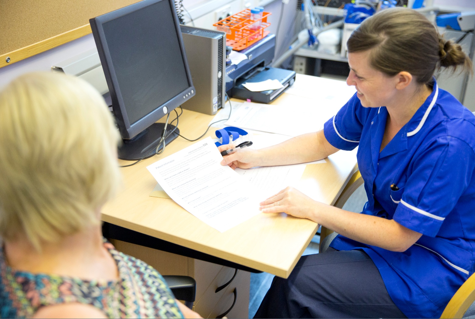
When we are recruiting for volunteers, we distribute flyers or posters with details of the study and how to contact us. For patient studies, we liaise with our clinical colleagues who help us to identify people who may want to take part. The clinician will describe the trial to the patient and ask if they would like to take part – if they are interested, one of the research team will describe the trial in more detail to them before asking them if they are willing to consent to being involved. Cambridge is a great city for clinical studies as we have a high rate of recruitment. We have many people who are motivated by research and they are interested and willing to give their time.
Why should people take part in research?
Research can potentially benefit a patient, for example if they are in a drug trial, but neither they nor the doctor know whether they will benefit before entering the trial. In my field of diagnostics imaging, we’re not usually giving patients a drug, and therefore there may be no direct benefit to the patient by entering the study. However, it could improve the care for future patients. Often patients are motivated simply by the chance to help someone in a similar position in the future.
How would you reassure people who are nervous at taking part in clinical research?
We are always very open with patients and explain clearly what will happen during the trial. All clinical trials are reviewed by an ethics committee before they are approved – they consider aspects of the trial that will directly affect the volunteers or patients who will be asked to take part. Patients are free to say they do not want to take part and do not have to give a reason for this – not taking part will not affect the treatment they were due to receive as part of normal care.
If people want to get involved in research, who should they contact?
In the first instance they should speak to the doctor in the hospital looking after them, the doctor usually has an idea of the trials that are open that they could be involved in. The hospital website also gives information about on-going trials and the National Institute for Health Research (NIHR) Cambridge BioResource is another approach that people can use to join medical research trials.
What would be your wish come true?
From a research point of view, it would be great if our work can directly change the lives of patients in the future. To answer your question another way, I really enjoy my job as there is something different every day – even if I won the lottery I wouldn’t give up my job!
Discover more about the NIHR Cambridge BRC
Contact us by phone, email or web for more information.

Events Calendar
Listing relevant events and training sessions for researchers and members of the public.